
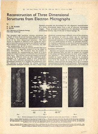
Optical filtering of electron micrographs + Reconstruction of three dimensional structures from electron micrographs
Publisher Information: 1966-1968.
Three-Dimensional Digital Image Processing
Rosier, D. J. de and Klug, Aaron (1926- ). (1) Optical filtering of electron micrographs: Reconstruction of one-sided images. Offprint from Nature 212 (1966). 8, [1]pp. 212 x 141 mm. Without wrappers as issued. (2) Reconstruction of three dimensional structures from electron micrographs. Offprint from Nature 217 (1968). 4, [1]pp. 277 x 205 mm. Without wrappers as issued. Together 2 offprints. Light toning, otherwise fine.
First Separate Editions. Klug and de Rosier invented methods for two-dimensional and three-dimensional digital image processing of electron microscope images. The latter method provided the theory behind the development of computed tomography (CT) by Hounsfield in the early 1970s. Hounsfield's invention of CT in the early 1970s benefited from several discussions with Klug about digital imaging (personal communication from Klug to J. Norman). Hounsfield received a share of the 1979 Nobel Prize in physiology/medicine for his invention of CT, which provided the first means for visualizing, in three dimensions, virtually all types of tissue within the body (see G-M 2700.4 and Grolier Medical Hundred 100). Klug received the Nobel Prize for chemistry in 1982 "for his development of crystallographic electron microscopy and his structural elucidation of biologically important nucleic acid-protein complexes." A press release written at the time refers to Klug's two- and three-dimensional imaging methods as an important part of his work: Electron microscopy has long been used to obtain a two-dimensional picture of biological objects. The power of the method to give a clear picture of the structure is, however, limited by several factors. The molecules of life consist mainly of light atoms, which makes the picture lacking in contrast. Increased contrast can be achieved with long exposure times, but this entails the danger that the structure is destroyed by radiation damage. Instead the contrast is generally improved by "staining" with heavy metals, which can also lead to a distortion of the structure. Klug has shown that pictures of biological objects seemingly lacking in contrast often contain a large amount of structural information, which can be made available by a mathematical manipulation of the original picture. His method allows electron microscope pictures of high quality to be obtained with very low radiation doses and without the use of heavy metal stains. In this way changes in the sample are minimized, so that the electron microscope picture at high resolution is a true representation of the original biological structure. The method gives a two-dimensional projection of the sample only, but Klug has shown that a three-dimensional reconstruction of the object can be obtained by collecting pictures in several different directions of projection (Nobel e-museum). 38691
Book Id: 38691Price: $4,750.00