
Cours de microscopie. Text and atlas
Publisher Information: Paris: Bailliere, 1845.
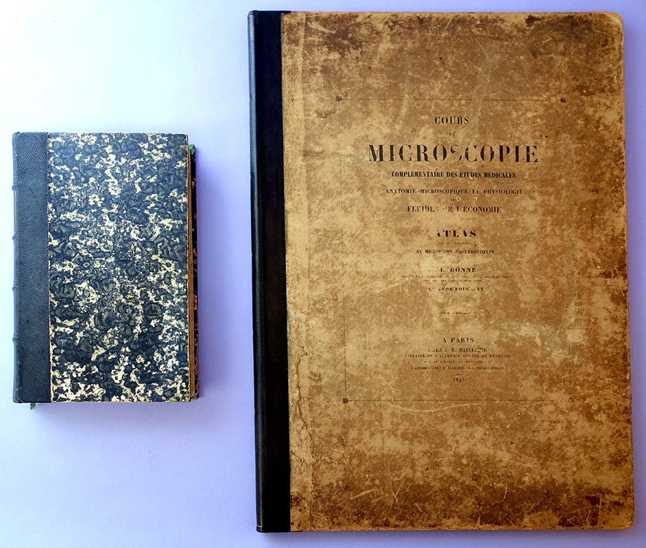
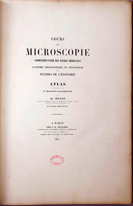
Donné, Alfred François (1801-78). Cours de microscopie complémentaire des études médicales . . . Text and atlas. [4], ii, [2, incl. errata], 550, pp. (text); 30pp. plus 20 plates engraved by Ouvret after micro-daguerreotypes taken by Léon Foucault (1819-68), original tissue guards present. Paris: J.-B. Baillière [etc.], 1844-45. 214 x 130 mm. (text); 436 x 300 mm. (atlas). Text in 19th century quarter morocco, mottled boards, light edgewear; atlas in original printed boards, new morocco spine, light rubbing and edgewear, a few minor stains; the two preserved in a cloth folding box. Minor foxing, text with library stamp on title and 3 or 4 other leaves, atlas with small library stamp on title and verso of each plate but very good overall.
First Edition of a major landmark in the fields of hematology, oncology, bacteriology, medical microscopy and photomicrography. Donné, a French public health physician, began teaching his pioneering course on medical microscopy in 1837, a time when the medical establishment remained largely unconvinced of the microscope’s usefulness as a diagnostic and investigative tool. In July 1839 Louis Daguerre, one of the inventors of photography, announced to the Académie des Sciences his “daguerreotype” process for creating finely detailed photographic images on specially prepared glass plates. Donné immediately embraced this new art and within a few months had created not only the first documented photographic portrait in Europe, but also the earliest method of preparing etched plates from daguerreotypes. Donné resolved to incorporate photography into his microscopy course, and in February 1840 he presented to the Académie his first photographic pictures of natural objects as seen through the microscope. “It was Alfred Donné who foresaw the helpful role that projections of microscopic pictures could play during lectures on micrography” (Dreyfus, p. 38).
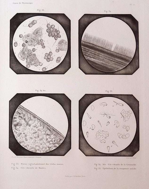
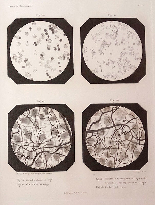
Over the next few years Donné continued to refine his photomicrography methods with the help of his assistant, Léon Foucault (who would go on to have a distinguished career as a physicist). In 1844 Donné published his Cours de microscopie complémentaire des études médicales (Course of microscopy complementary to medical studies), following it a year later with an atlas illustrated with 86 engravings copied from micro-daguerreotypes taken by Foucault. This extraordinary work was the first biomedical textbook to be illustrated with images made from photomicrographs. Among its noteworthy images are the first microphotographs of human blood cells and platelets, and the first photographic illustration of Trichomonas vaginalis, the protozoon responsible for vaginal infections, which Donné had discovered in 1836. The text volume of the Cours contains the first description of the microscopic appearance of leukemia, which Donné had observed in blood taken from both an autopsy and a living patient. His observations mark the first time that leukemia was linked with abnormal blood pathology:
"There are conditions in which white cells seem to be in excess in the blood. I found this fact so many times, it is so evident in certain patients, that I cannot conceive the slightest doubt in this regard. One can find in some patients such a great number of these cells that even the least experienced observer is greatly impressed. I had an opportunity of seeing these in a patient under Dr. Rayer at the Hôpital de la Charité. . . . The blood of this patient showed such a number of white cells that I thought his blood was mixed with pus, but in the end, I was able to observe a clear-cut difference between these cells, and the white cells . . . " (p. 135; translation from Thorburn, pp. 379-80).
The following year this abnormal blood condition was recognized as a new disease by both John Hughes Bennett (a former student of Donné’s) and Rudolph Virchow. Garrison-Morton.com 267.1, 3060.1. Dreyfus, Some Milestones in the History of Hematology, pp. 38-40, 54-56, 76-78. Frizot, A New History of Photography, p. 275. Gernsheim & Gernsheim, The History of Photography 1685-1914, pp. 116, 539. Hannavy, Encyclopedia of Nineteenth-Century Photography, Vol. 1, p. 1120. Wintrobe, Hematology: The Blossoming of a Science, p. 12. Bernard, Histoire illustrée de l’hématologie, passim. Thorburn, “Alfred François Donné, 1801-1878, discoverer of Trichomonas vaginalis and of leukaemia,” British Journal of Venereal Disease 50 (1974): 377-380.
Book Id: 35134Price: $15,000.00